News from the Field


Scientists Find Brain Pathways for Threat Response in Mice
Scientists at the National Institute on Alcohol Abuse and Alcoholism (NIAAA) have identified brain pathways that may coordinate an animal’s response to potentially traumatic situations. Understanding where and how neural circuits mediate such functions—and how they could malfunction—may provide clues about their role in trauma-related and stress-related psychiatric disorders in people. A report of this NIAAA research was published in the journal Nature.
“Experiencing or witnessing traumatic events is often at the root of trauma-related and stress-related psychiatric conditions including alcohol use disorder,” said the study’s senior author, Andrew Holmes, Ph.D., senior investigator in NIAAA’s Laboratory of Behavioral and Genomic Neuroscience. “In animal models of stress and trauma, learning about potential sources of threat by observing how others deal with danger can be an effective way to avoid harm. Understanding the differences in how the brain processes direct experience of a threat compared to observing another’s response to a threat may shed light on factors that predispose humans to trauma- and stress-related psychiatric disorders.”
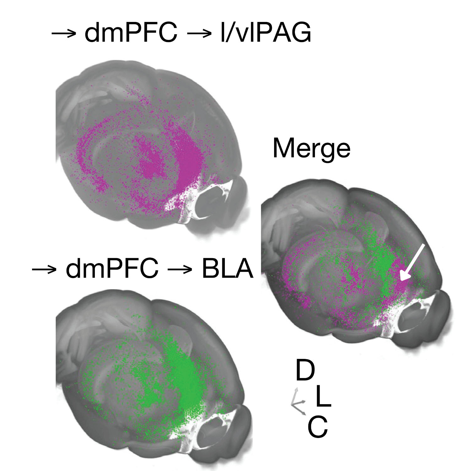
In their study, Dr. Holmes and his colleagues examined the neural basis of observational fear learning (OFL) in mice. OFL is the process through which animals learn about sources of danger and minimize their own risk by observing how others respond to threats. The researchers focused on an area at the front of the brain called the dorsomedial prefrontal cortex (dmPFC), which is known to play key roles in processing social information and interpreting threats in mice, humans, and other animals. The researchers found that the dmPFC is recruited and required for OFL in mice.
Then the researchers measured activity of neural pathways leading to and away from the dmPFC in mice that watched other mice learn to associate a sound cue with a mild foot shock. Animals that receive this cue-shock pairing typically learn to “freeze” or become motionless when they hear the sound cue. The scientists then presented the observer mice with the sound cue-foot shock pairing and measured activity in the same dmPFC neural pathways.
The researchers found that when observer mice faced the “threat” of the sound cue, they showed a coordinated activation of pathways that either mobilized or suppressed the freezing response. For example, the observer mice showed that dmPFC projections to the midbrain periaqueductal gray (PAG) regulate OFL and that amygdalar and hippocampal inputs to the dmPFC opposingly modulate observer freezing. Hippocampal inputs constrained, whereas basolateral amygdala inputs promoted freezing in observer mice.
The researchers hypothesized that a critical function of the dmPFC in the observer mice may be to balance the need to minimize harm (i.e., freezing) with the need to fulfill other essential survival functions (e.g., assessing risk or comforting others).
The researchers also said that the findings suggest that maladaptive responses to socially learned threats could arise in part from deficits in the dmPFC pathways and may point to a potential role of the dmPFC deficits in trauma- and stress-related psychiatric disorders in humans.
Reference:
Silverstein SE, O’Sullivan R, Bukalo O, Pati D, Schaffer JA, Limoges A, Zsembik L, Yoshida T, O’Malley JJ, Paletzki RF, Lieberman AG, Nonaka M, Deisseroth K, Gerfen CR, Penzo MA, Kash TL, Holmes A. A distinct cortical code for socially learned threat. Nature. 2024 Feb;626(8001):1066-1072. PubMed PMID: 38326610